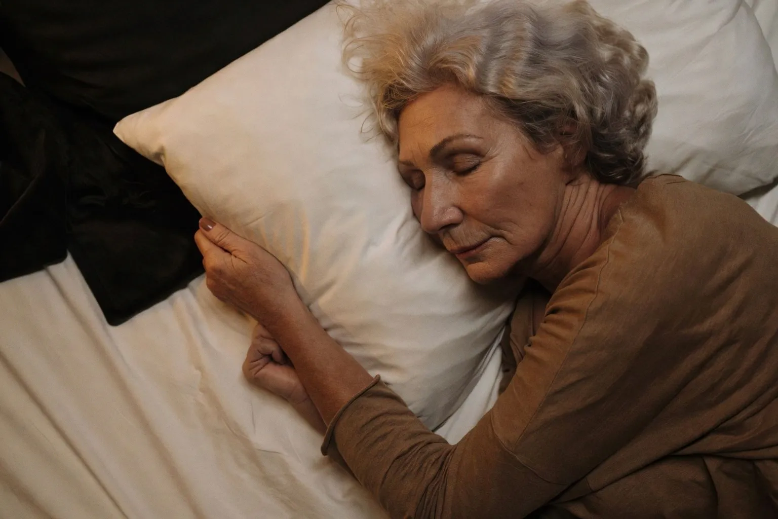
A team from Binghamton University analyzed resting-state fMRI scans from over 1,300 adults and found that poor sleep quality remodels brain network connectivity in fundamentally different ways depending on age — with the pattern in older adults closely resembling the earliest silent stages of Alzheimer's disease. The study, published in Neurobiology of Aging, is one of the first to map how sleep disturbance separates into distinct neural signatures across the adult lifespan. For anyone tracking cognitive decline biomarkers, this shifts the conversation from "how many hours" to which specific network dysregulation is active and when.
Two Ages, Two Mechanisms
In college-aged adults reporting poor sleep, the researchers observed overconnectivity in brain regions governing physical movement — motor and premotor networks firing at a baseline level inconsistent with sleep onset. The interpretation is straightforward: the young, poorly sleeping brain is not physically ready to rest. The motor system remains in a state of activation, suggesting physiological hyperarousal that precedes the cognitive complaint.
In adults aged 65 and older, the picture inverts. Movement-related regions are underconnected. Instead, the dominant pattern is hyperconnectivity within and between cognitive networks — specifically the Default Mode Network (DMN), which handles internal thought and memory retrieval, and the Frontal Parietal Network (FPN), responsible for sustained attention and working memory. This isn't the same mechanism as in younger brains; it's a systemic breakdown in sleep-regulation architecture.
The DMN–FPN Hyperconnectivity Signal
The most clinically significant finding centers on older women. In this subgroup, abnormal overcommunication between the DMN and FPN was directly associated with poorer memory performance. This same connectivity pattern — excessive cross-talk between internally focused and externally directed networks — is a recognized feature of preclinical Alzheimer's pathology. The researchers noted this mirrors wiring configurations observed in the silent, asymptomatic stages of the disease, years before clinical diagnosis.
The causal direction remains unresolved: does dysfunctional connectivity produce poor sleep, or does chronic sleep disruption drive the network abnormality? Growing evidence, however, points to between-network hyperconnectivity — particularly involving the DMN — as an early marker of declining brain health rather than a benign correlate. Cognitive consequences appear to follow sleep disturbance or the associated connectivity shifts over time.
Potential Mechanisms and Open Variables
The researchers flagged several explanatory hypotheses. Older adults may habituate to hyperarousal states, developing coping behaviors — including increased willingness to use sleep medications — that mask the underlying dysregulation. Rumination, defined as repetitive overthinking often comorbid with anxiety and depression, is another candidate: pre-sleep cognitive agitation prevents the calm physiological state required for onset. The depression–dementia link is relevant here, with some evidence that depressive states can mimic cognitive decline, though cognition may improve with depression treatment. Whether these factors are independent contributors or overlapping manifestations of the same neural disruption is not yet established.
Biological sex emerged as a meaningful variable, with older women showing the most pronounced DMN–FPN hyperconnectivity. The study did not isolate hormonal or mechanistic explanations for this sex difference, but the signal is consistent with broader research on sex-specific vulnerability in neurodegeneration.
The protocol-level takeaway is precise: age-stratified sleep assessment should account for which networks are dysregulated, not just subjective sleep quality scores. A 22-year-old with motor hyperconnectivity and a 68-year-old with DMN–FPN overcommunication are presenting different neural problems with potentially different intervention targets — and different long-term risk profiles.