Your brain operates at a voltage roughly one-millionth that of a wall outlet. A typical electroencephalogram captures cortical oscillations between 10 and 100 microvolts — a signal so faint that the electrical buzz of nearby wiring, the flicker of an eyelid, or a clench of the jaw can bury it entirely. When a consumer headband streams data to your phone, what you are looking at is not raw neural output. It is a composite of cortical activity, muscle tension, power-grid leakage, and electrode noise — all compressed, filtered, and rendered in a graph that rarely labels which is which.
This matters because the frequency bands most useful for performance tracking — beta (13–30 Hz) and gamma (above 30 Hz) — overlap almost perfectly with the spectral footprint of electromyographic contamination from the temporalis and frontalis muscles. If you cannot distinguish signal from artifact, you are not measuring cognition. You are measuring the tension in your jaw.
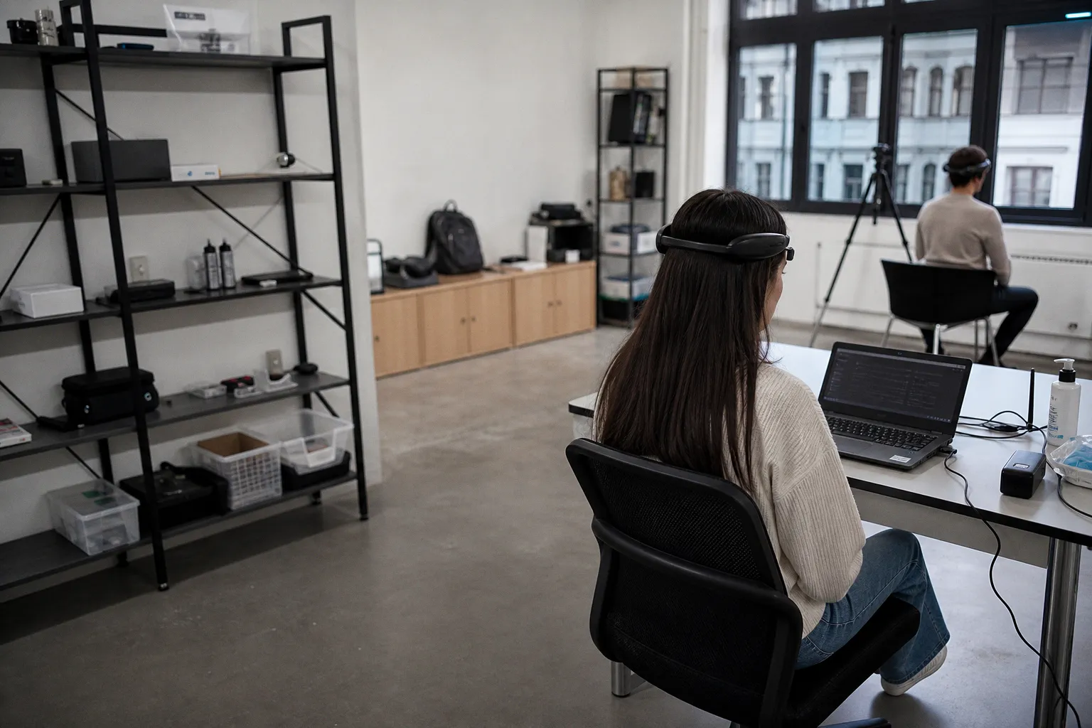
The Microvolt Challenge: Why Consumer Dry Electrodes Invite Noise
Every EEG system faces the same fundamental problem: the signal it seeks is orders of magnitude weaker than the electromagnetic environment it lives in. Clinical-grade setups mitigate this with wet-gel electrodes that form a low-impedance bridge between scalp and sensor, minimizing contact resistance to under 5 kilohms. Consumer headbands like the Muse 2 or Emotiv Epoc X use dry electrodes — polymer or spring-loaded contacts pressed against the skin with no conductive gel.
The trade-off is convenience versus signal integrity. Dry electrodes produce higher contact impedance, often exceeding 50 kOhm, which creates two compounding failure modes:
- Motion sensitivity. Even minor shifts in head position break the skin-electrode interface, generating voltage transients that look like sharp neural events in the time domain but carry zero cortical information.
- Higher noise floor. Elevated impedance amplifies common-mode interference — the ambient electromagnetic radiation from power lines, monitors, and fluorescent lighting — that a low-impedance electrode would shunt to ground.
The result is a data stream with a fundamentally worse signal-to-noise ratio before any digital filtering is applied. The headband's onboard firmware may smooth this with aggressive band-pass filtering (typically 0.5–40 Hz), but aggressive filtering introduces its own distortions: phase shifts near the filter edges and attenuation of genuine high-frequency neural content.
If your EEG headband promises "lab-grade brainwave data" from dry electrodes sitting on your forehead, the burden of proof is on the hardware — and the microvolt-level signal is already losing before the first filter runs.
Understanding this baseline fragility is step one. You cannot audit what you do not understand the constraints of.
Spectral Ghosts: Identifying 50 Hz and 60 Hz Power Line Interference
In any EEG frequency-domain analysis, the single most recognizable artifact is the sharp, narrow spectral peak at your local mains frequency: 50 Hz in Europe and Asia, 60 Hz in North America. It appears as an isolated spike in the power spectrum that has no neurophysiological origin whatsoever — it is electromagnetic leakage coupling into the electrode leads.
The standard remediation is a notch filter, a digital trap that attenuates energy at exactly that frequency. But a notch filter is not surgical:
- It removes a band of frequencies centered on the target, not just the single line. A 50 Hz notch with even a moderate Q-factor will also suppress legitimate neural oscillations in the 48–52 Hz range — which, for research-grade gamma analysis, is a meaningful loss.
- It does not eliminate harmonics. Power line interference often presents at integer multiples (100 Hz, 150 Hz, 200 Hz for 50 Hz mains). Consumer headband firmware rarely addresses harmonics, so residual contamination leaks into lower-frequency bands through aliasing if the sampling rate is inadequate.
How to spot it in your own data: Compute a fast Fourier transform (FFT) of a 10-second resting-state window. A clean neural spectrum shows a smooth 1/f distribution — power declining with increasing frequency. If you see a single, unnaturally sharp peak at exactly 50 or 60 Hz with a width of less than 1 Hz, that is mains noise, not a cortical oscillation. No brain region generates a perfectly stable single-frequency signal.
Ocular and Muscle Contaminants: Decoding Blinks and EMG Masking
Two artifact classes dominate the time domain: eye blinks and muscle tension. They are not subtle — but in a consumer device's simplified visualization, they are routinely mistaken for cognitive events.
Eye blinks produce high-amplitude, low-frequency deflections. The eyeball acts as a dipole (the cornea is electrically positive relative to the retina), and a blink causes this dipole to rotate — a phenomenon known as Bell's Phenomenon. The resulting artifact peaks at 100 microvolts or more, far exceeding any cortical signal. It appears predominantly in frontal electrodes (Fp1, Fp2) and occupies the 0.5–4 Hz range — the delta and theta bands. A consumer device placed on the forehead sits directly above the orbital muscles, making it maximally susceptible.
| Artifact Type | Amplitude | Frequency Range | Primary Electrode Sites | Confusion Risk |
|---|---|---|---|---|
| Eye blink | >100 µV | 0.5–4 Hz | Fp1, Fp2 (frontal) | Delta/theta "deep focus" readings |
| Jaw clench (EMG) | 50–500 µV | 20–500 Hz broadband | Temporal, frontal | Beta/gamma "concentration" scores |
| Neck tension (EMG) | 20–200 µV | 13–100 Hz | Occipital, parietal | Beta-band "active thinking" |
| Electrode pop | Variable, transient | Broadband, single spike | Any channel | False event markers |
Muscle artifacts are more insidious. Jaw clenching, frowning, and even sustained talking generate electromyographic signals that are broadband and high-frequency — they blanket the beta and gamma ranges entirely. A frontalis muscle contraction at 30 µV of EMG energy can sit directly on top of a 5 µV beta oscillation, effectively doubling the apparent "beta power" in a spectral analysis. Consumer platforms that report "focus scores" derived from beta-band activity may be measuring facial tension, not attentional engagement.
A 30 µV muscle artifact sitting on a 5 µV beta oscillation doubles apparent focus-level readings. Your concentration metric may just be your jaw.
The practical test: clench your jaw for five seconds during a recording, then relax. If your headband's reported beta power drops noticeably in the relaxation window, your baseline readings are contaminated by tonic muscle activity, and every "focus" session logged without jaw-position awareness is suspect.
Electrode Pops and Impedance Drift: Detecting Physical Signal Breaks
Beyond biological artifacts, the electrode interface itself introduces noise. An "electrode pop" is a sudden, sharp voltage jump — sometimes exceeding several hundred microvolts — followed by a slow exponential drift back to baseline. The cause is an abrupt change in skin-electrode impedance: the electrode shifts position, a hair slides underneath, or the user's skin conductance changes due to perspiration.
In time-domain plots, pops appear as isolated vertical spikes that look superficially like high-amplitude neural events. In the frequency domain, they scatter broadband energy across the entire spectrum, creating a transient elevation in apparent power at every frequency band simultaneously. If your analysis pipeline counts "events" above a power threshold, electrode pops will inflate your event count — sometimes dramatically.
Impedance drift is slower and less visible. Over a 30-minute session, skin temperature rises, sweat composition changes, and the electrode-skin interface degrades. Contact impedance may start at 30 kOhm and climb to 100+ kOhm. The practical effect is a gradual increase in the noise floor and a slow, invisible loss of signal fidelity. The data still arrives; it is simply less trustworthy than it was at minute one.
Validating the Stream: Sampling Rates and the Research-Grade Baseline
The Nyquist-Shannon theorem mandates that a sampling rate must be at least twice the highest frequency of interest. For beta-band analysis (up to 30 Hz), a minimum of 60 samples per second is mathematically required. For gamma-band content up to 40 Hz, 80 Hz sampling suffices in theory. In practice, research-grade systems sample at 250 Hz or higher to allow for anti-aliasing filters that roll off gradually without distorting the passband.
Most consumer headbands sample between 250 Hz and 256 Hz — nominally adequate. The problem is not the raw rate but what happens before the data reaches your application. Onboard processors in consumer devices apply real-time filtering, compression, and sometimes proprietary "denoising" algorithms that are opaque to the end user. By the time the data stream arrives on your phone, it has been transformed in ways you cannot fully reverse or audit.
A minimum validation protocol for any consumer EEG stream:
1. Visual inspection of the raw time-domain signal. Look for amplitude excursions above 100 µV (likely blinks or pops) and sustained rhythmic patterns that do not correspond to known brainwave frequencies (likely 50/60 Hz leakage or EMG).
2. Spectral decomposition via FFT. Compute the power spectrum of a 10–20 second resting window. Check for the mains-frequency spike, broadband elevation in the 20–50 Hz range (EMG), and the absence of a clean alpha peak at 8–12 Hz during eyes-closed recording (which would indicate the device is failing to capture cortical oscillations at all).
3. Blink-corruption test. Perform deliberate blinks at known intervals and check whether the time-domain trace shows corresponding >100 µV deflections in frontal channels. If blinks produce no visible artifact, the device's firmware is filtering aggressively enough to potentially remove legitimate low-frequency neural data along with the blink.
4. EMG contamination test. Clench and release the jaw during recording. A clean signal should show minimal beta-band power change. A spike in beta/gamma power during clenching confirms that muscle artifacts are masquerading as neural activity.
5. Session-over-session drift comparison. Record two 5-minute resting-state sessions 30 minutes apart. Compute the power spectrum for each. If the noise floor rises by more than 10–15 dB in the second session, impedance drift is degrading data quality over time, and long-session recordings are progressively unreliable.
These checks take under 20 minutes. They will not transform a consumer device into a clinical system, but they will tell you exactly how much of your reported brainwave data is actually brain-derived — and how much is the electrical environment, your own muscles, and the electrode sitting on your skin.
The consumer neurotech market moves fast, and the gap between marketing language and signal integrity remains wide. Devices sold for meditation tracking, sleep scoring, and "focus optimization" are delivering data streams that require the same artifact literacy as a research EEG — minus the training, minus the wet electrodes, and minus the transparency about what the onboard firmware has already done to your signal before you ever see it. Treat the output as a hypothesis, not a measurement, until you have audited it yourself.